40 brain mri with labels
Automated MRI image labelling processes 100,000 brain exams in under 30 ... Researchers from the School of Biomedical Engineering & Imaging Sciences at King's College London have automated brain MRI image labeling, needed to teach machine learning image recognition models,... › en › e-AnatomyShoulder: MRI, radiographical, and illustrated anatomical ... Sep 13, 2021 · MRI of the shoulder : muscles of the rotator cuff labeled on a sagittal MR slice. An MRI of the shoulder of a healthy subject was performed in the 3 planes of space (coronal, axial, sagittal) commonly used in osteoarticular imaging, with two weightings to explore the musculoskeletal pathology of the shoulder: spin-echo T1 and proton-density ...
Magnetic resonance imaging of the brain - Wikipedia History. The first MR images of a human brain were obtained in 1978 by two groups of researchers at EMI Laboratories led by Ian Robert Young and Hugh Clow. In 1986, Charles L. Dumoulin and Howard R. Hart at General Electric developed MR angiography, and Denis Le Bihan obtained the first images and later patented diffusion MRI. In 1988, Arno Villringer and colleagues demonstrated that ...

Brain mri with labels
Brain: Atlas of human anatomy with MRI - e-Anatomy - IMAIOS MRI Atlas of the Brain. This page presents a comprehensive series of labeled axial, sagittal and coronal images from a normal human brain magnetic resonance imaging exam. This MRI brain cross-sectional anatomy tool serves as a reference atlas to guide radiologists and researchers in the accurate identification of the brain structures. Cross-sectional anatomy of the brain - e-Anatomy - IMAIOS We created a brain atlas that is an interactive tool for studying the conventional anatomy of the normal brain based on a magnetic resonance imaging exam of the axial brain. Anatomical structures and specific areas are visible as interactive labeled images. Cross sectional anatomy: MRI of the brain. An MRI was performed on a healthy subject ... › pmc › articlesPolitical Orientations Are Correlated with Brain Structure in ... Apr 26, 2011 · To test the hypothesis that political liberalism (versus conservatism) is associated with differences in gray matter volume in anterior cingulate cortex, we recorded structural magnetic resonance imaging (MRI) scans from 90 healthy young adults (61% female) who self-reported their political attitudes confidentially on a five-point scale from ...
Brain mri with labels. A normative spatiotemporal MRI atlas of the fetal brain for automatic ... through manual segmentation the following structures (right and left, when applicable) were labeled on fetal brain mri atlases: hippocampi, amygdala, fornix, cerebellum, brainstem, caudate nuclei, thalami, subthalamic nuclei, lentiform nuclei, corpus callosum, lateral ventricles, developing white matter, cortical plate, and cerebrospinal fluid … Labeled MRI Brain Scans - Neuromorphometrics We can also label scans that you provide and we are very interested in labeling white matter anatomy as seen in diffusion-weighted MRI scans. If you want an aggregate version of our data, we can provide it as a probabilistic atlas. The cost to label a single scan is $2449 (USD). Clinical and Arterial Spin Labeling Brain MRI Features of Transitional ... We investigate whether arterial spin labeling (ASL) magnetic resonance imaging (MRI) can distinguish brain TVAs from DVAs and guide their clinical management. Methods: We conducted a single-center retrospective review of patients with brain parenchymal DVA-like lesions with increased ASL signal on MRI. Clinical histories and follow-up ... : Automated MRI Brain volumetry system It is intended to help researchers all over the world to obtain automatically volumetric brain information from their MRI data without the need for any infrastructure in their local sites. volBrain works in a fully automatic manner and is able to provide brain structure volumes without any human interaction.
brain anatomy | MRI coronal brain anatomy | free MRI cross sectional ... This MRI brain coronal cross sectional anatomy tool is absolutely free to use. Use the mouse scroll wheel to move the images up and down alternatively use the tiny arrows (>>) on both side of the image to move the images.>>) on both side of the image to move the images. › AANLIB › casesHarvard University Show labels Show list All modalities to: MR-T1 MR-T2 FDG T1/FDG T2/FDG Deep learning to automate the labelling of head MRI datasets for ... In order to generate 'reference-standard image labels' for model testing, 950 head MRI examinations were randomly selected from the 5000 examinations with reference-standard report labels. Two neuroradiologists labelled 250 examinations as normal or abnormal applying the same framework used for report labelling—but interrogating the ... Brain MRI: How to read MRI brain scan | Kenhub MRI is the most sensitive imaging method when it comes to examining the structure of the brain and spinal cord. It works by exciting the tissue hydrogen protons, which in turn emit electromagnetic signals back to the MRI machine. The MRI machine detects their intensity and translates it into a gray-scale MRI image.
Atlas of BRAIN MRI - W-Radiology Brain magnetic resonance imaging (MRI) is a common medical imaging method that allows clinicians to examine the brain's anatomy (1). It uses a magnetic field and radio waves to produce detailed images of the brain and the brainstem to detect various conditions (2). Automatic anatomical brain MRI segmentation combining label propagation ... Using the brain label data, we determined the variation of a and b depending on the registration method employed (unregistered, ... Manual and automated measurement of the whole thalamus and mediodorsal nucleus using magnetic resonance imaging. NeuroImage, 17 (2) (2002), pp. 631-642. Article Download PDF View Record in Scopus Google Scholar. Automatic Structural Parcellation of Mouse Brain MRI Using Multi-Atlas ... In this paper, we apply a fully automatic multi-atlas based open source framework to the structural parcellation of mouse brain MRI. The framework consist of several preprocessing steps along with a non-rigid B-spline parameterised registration [31], and the above mentioned label fusion method STEPS. UCLA Brain Mapping Center - ICBM Template To view both the structural MRI and the labels launch the program typing Display icbm_template.mnc -label icbm_labels_corrected.mnc. The opacity of the labels can be set in the Colour Coding menu. The number of each label appears at the bottom left of the orthogonal views window.
Diagnostic Imaging - Brain MRI
Labels · Akriti3/Brain_MRI_using_Pytorch · GitHub Contribute to Akriti3/Brain_MRI_using_Pytorch development by creating an account on GitHub.
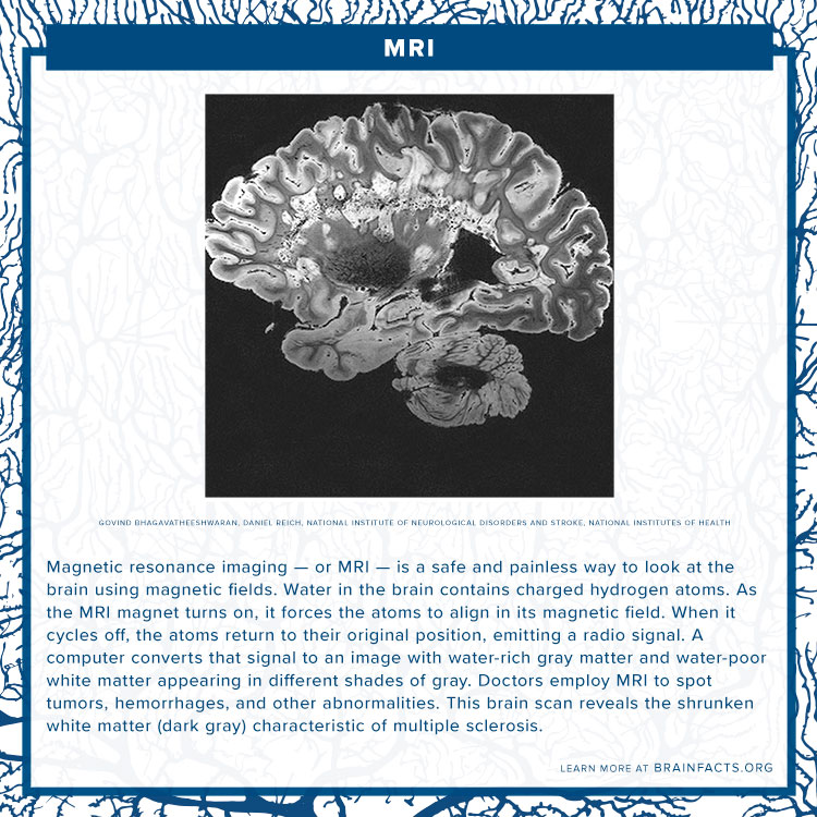
MRI
› 2013 › 07CPT Code for MRI Brain, Breast, Lumbar Spine and Shoulder Find below the latest Radiology CPT codes for for MRI of Brain, Breast, Lumbar Spine and Shoulder: CPT Codes for MRI Lumbar spine In human Lumbar spine is represented by the 5 vertebrae in between the ribcage and the pelvis forming the largest segment of the vertebral column. Depending on the condition that one is treated on these parts of the ...

Neuroradiology Cases: Multiple System Atrophy MRI
MRI Brain Animated Quiz - University of Minnesota Note: spacebar toggles labels; also arrow keys do Previous/Next Sequentially click/tap: first the dot associated with a term; then, its corresponding target dot on the MRI image. If a line connection appears, your choice was correct!

Head Case: What this about your brain, now?
What Does a Brain MRI Show? - San Diego Health What does a brain MRI show? The answer is, unfortunately, not very. MRI scans (magnetic resonance imaging) have been around for decades, and the technology has been steadily improving. Today, a brain MRI test can identify whether or not a person has a stroke, or if the person has suffered a traumatic brain injury, or if the person is suffering ...
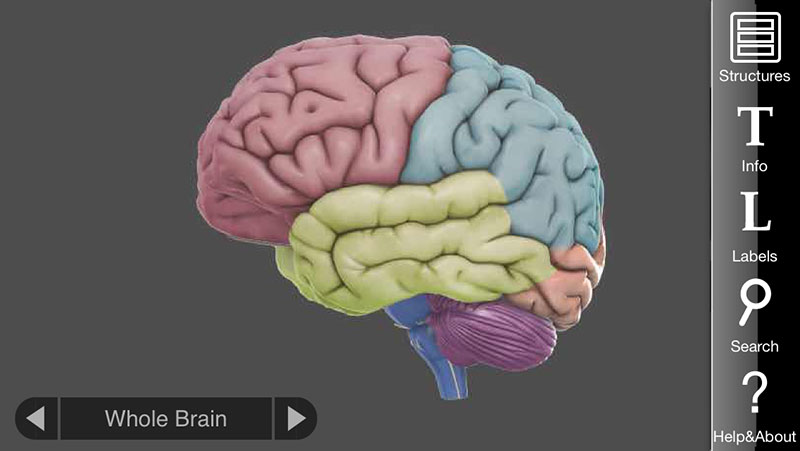
3D Brain App, an interactive way to learn about the different parts of the human brain | Apps ...
Brain MRI Dataset | Kaggle Brain MRI Dataset | Kaggle. View Active Events. Haşim Mumcu · Updated 3 years ago. arrow_drop_up. 4. New Notebook. file_download Download (8 MB) more_vert.

Radiology MRI: Chronic Subdural Hematoma
Researchers automate brain MRI image labelling, more than 100,000 exams ... July 22, 2021 King's College London Researchers from the School of Biomedical Engineering & Imaging Sciences have automated brain MRI image labelling, needed to teach machine learning image recognition models, by deriving important labels from radiology reports and accurately assigning them to the corresponding MRI examinations.

Dr Balaji Anvekar FRCR: Frontal subcortical white matter cystic lesions MRI
analyticsindiamag.com › brain-tumor-predictionBrain Tumor Prediction Through MRI Images Using CNN In Keras Aug 19, 2020 · Both the folders contain different MRI images of the patients. Yes folder has patients that have brain tumors whereas No folder has MRI images of patients with no brain tumor. There are a total of 155 images of positive patients of brain tumor and 98 images of other patients having no brain tumor. All the images are of 240X240 pixels.

ventricular system overview :: Brain Imaging
Labeled imaging anatomy cases | Radiology Reference Article ... This article lists a series of labeled imaging anatomy cases by body region and modality. Brain CT head: non-contrast axial CT head: non-contrast coronal CT head: non-contrast sagittal CT head: angiogram axial CT head: angiogram coronal CT...

Brain Radiology | 118.164
Frontiers | 101 Labeled Brain Images and a Consistent Human Cortical ... Labeled anatomical subdivisions of the brain enable one to quantify and report brain imaging data within brain regions, which is routinely done for functional, diffusion, and structural magnetic resonance images (f/d/MRI) and positron emission tomography data.

Brain Radiology | 119.164
Researchers automate brain MRI image labeling, more than 100,000 exams ... Researchers have automated brain MRI image labeling, needed to teach machine learning image recognition models, by deriving important labels from radiology reports and accurately assigning them to...

Neuroanatomy - encyclopedia article - Citizendium
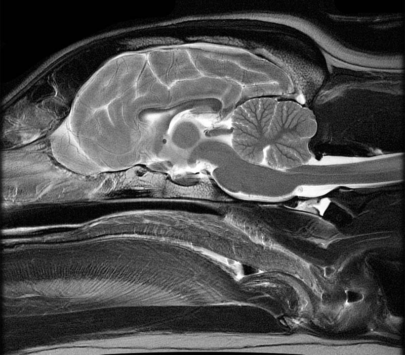
MRI Brain Atlas This web app Atlas is intended for veterinary students and radiologists seeking quick access to canine brain anatomy through a mobile device. Via a toggle button, either MRI images or approximately comparable Brain Transection images may be viewed with or without labels. Navigation & Labels.
![Untitled Document [www.swjpcc.com]](http://static1.1.sqspcdn.com/static/f/654826/26424684/1438174189687/100-15+Figure+3.gif?token=VZCSSTefJcURwXVEtWCFxzi8s2Y%3D)
Untitled Document [www.swjpcc.com]
NITRC: Manually Labeled MRI Brain Scan Database: Tool/Resource Info This is a continuously growing and improving database of high-quality neuroanatomically labeled MRI brain scans, created not by an algorithm, but by neuroanatomical experts. All results are checked and corrected. Regions of interest include the usual sub-cortical structures (thalamus, caudate, putamen, hippocampus, etc), along with ventricles ...

Dr Balaji Anvekar FRCR: Hypothalamic Hamartoma presenting with precocious puberty
MRI anatomy | free MRI axial brain anatomy MRI anatomy | free MRI axial brain anatomy This MRI brain cross sectional anatomy tool is absolutely free to use. Use the mouse scroll wheel to move the images up and down alternatively use the tiny arrows (>>) on both side of the image to move the images.

356 best MRI images on Pinterest | Radiology, Rad tech and Anatomy
CaseStacks.com - MRI Brain Anatomy Labeled scrollable brain MRI covering anatomy with a level of detail appropriate for medical students. Show/Hide Labels. MRI Brain Anatomy. Back to Anatomy overview. Facebook; Twitter; ... Labelled radiographs and CT/MRI series teaching anatomy with a level of detail appropriate for medical students and junior residents. Pelvis. Pelvic MRI anatomy
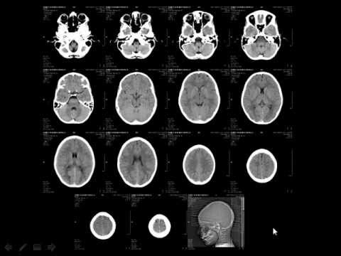
Radiology - Normal brain anatomy - CT and MRI - YouTube
Brain lobes - annotated MRI | Radiology Case | Radiopaedia.org Labeled Anatomy by Abdullah Abohimed; Brain tracts on mri by Khurshed Abdujabborov; theangrydoctor by Dr Mudit Arora; UOE MB2 Neuroanatomy P1 S4 by UoE Radiology; Annotated Anatomy by Marc Hidalgo; NRAD by Johann Jende; Neuroanatomy by Dr. Rouslan Senkeev; Brain Anatomy MRI by R. Furman Borst MD; Annotated CT/MR Teaching by Matt Wong Neuro- MRI ...
Sagittal Plane MRI Head Atlass
en.wikipedia.org › wiki › Diffusion_MRIDiffusion MRI - Wikipedia Diffusion-weighted magnetic resonance imaging (DWI or DW-MRI) is the use of specific MRI sequences as well as software that generates images from the resulting data that uses the diffusion of water molecules to generate contrast in MR images.
Post a Comment for "40 brain mri with labels"