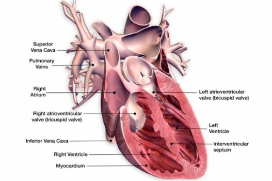
43 the human heart and its labels
Diagram of Human Heart and Blood Circulation in It Exterior of the Human Heart A heart diagram labeled will provide plenty of information about the structure of your heart, including the wall of your heart. The wall of the heart has three different layers, such as the Myocardium, the Epicardium, and the Endocardium. Here's more about these three layers. Epicardium File:Diagram of the human heart (no labels).svg - Wikimedia File:Diagram of the human heart (no labels).svg. From Wikimedia Commons, the free media repository. File. File history. File usage on Commons. Metadata. Size of this PNG preview of this SVG file: 498 × 599 pixels. Other resolutions: 199 × 240 pixels | 399 × 480 pixels | 639 × 768 pixels | 851 × 1,024 pixels | 1,703 × 2,048 pixels | 533 × ...
The Human Heart Labeling Worksheet (Teacher-Made) - Twinkl The human heart is a muscle made up of four chambers, these are: Two upper chambers - the left atrium and right atrium Two lower chambers - the left and right ventricles. It's also made up of four valves - these are known as the tricuspid, pulmonary, mitral and aortic valves.

The human heart and its labels
A Diagram of the Heart and Its Functioning Explained in Detail The heart blood flow diagram (flowchart) given below will help you to understand the pathway of blood through the heart.Initial five points denotes impure or deoxygenated blood and the last five points denotes pure or oxygenated blood. 1.Different Parts of the Body ↓ 2.Major Veins ↓ 3.Right Atrium ↓ 4.Right Ventricle ↓ 5.Pulmonary Artery ↓ 6.Lungs Labelling the heart — Science Learning Hub Labelling the heart — Science Learning Hub Labelling the heart Add to collection The heart is a muscular organ that pumps blood through the blood vessels of the circulatory system. Blood transports oxygen and nutrients to the body. It is also involved in the removal of metabolic wastes. Topics Concepts Citizen science Teacher PLD Glossary Sign in Heart Diagram with Labels and Detailed Explanation - BYJUS Diagram of Heart. The human heart is the most crucial organ of the human body. It pumps blood from the heart to different parts of the body and back to the heart. The most common heart attack symptoms or warning signs are chest pain, breathlessness, nausea, sweating etc. The diagram of heart is beneficial for Class 10 and 12 and is frequently ...
The human heart and its labels. Simple Heart Diagram Labeling Activity (Teacher-Made) - Twinkl This simple heart diagram with labels is a fab learning activity to help your pupils aged 10-11 understand the heart and its function in the human body. ... The resource comes with two different diagrams of the heart; one with labels attached, and one blank diagram with the labels at the bottom for students to complete themselves. Ideal as an ... How to Draw and Label Human Heart || Easiest Method || Learn Biology ... #humanheart #biology #drawandlabel #drawing #sumairaanwar › photos › male-human-anatomyMale Human Anatomy Diagram Pictures, Images and Stock Photos Human internal organs Internal organs in woman and man body. Brain, stomach, heart, kidney, medical icon in female and male silhouette. Digestive, respiratory, cardiovascular systems. Anatomy poster vector illustration. male human anatomy diagram stock illustrations A Labeled Diagram of the Human Heart You Really Need to See The human heart, comprises four chambers: right atrium, left atrium, right ventricle and left ventricle. The two upper chambers are called the left and the right atria, and the two lower chambers are known as the left and the right ventricles. The two atria and ventricles are separated from each other by a muscle wall called 'septum'.
developer.apple.com › design › human-interfaceDesigning for watchOS - Platforms - Human Interface ... People also appreciate taking advantage of data that device features — like GPS, sensors for blood oxygen and heart function, altimeter, accelerometer, and gyroscope — can provide. App interactions. How to Draw a Human Heart: 11 Steps (with Pictures) - wikiHow Label the parts of the heart if you'd to reference it for anatomy. If you're trying to identify parts of the heart for a class you're taking, it's good practice to draw the heart yourself and label each segment. You can refer to your textbook in order to label the: [9] Aorta Superior vena cava Inferior vena cava Right and left atria Human Heart Diagram Labeled | Science Trends Human Heart Diagram Labeled Daniel Nelson 1, January 2019 | Last Updated: 3, March 2020 The human heart is an organ responsible for pumping blood through the body, moving the blood (which carries valuable oxygen) to all the tissues in the body. Without the heart, the tissues couldn't get the oxygen they need and would die. Human Heart Labeling Teaching Resources | Teachers Pay Teachers Human Heart Parts and Blood Flow Labeling Worksheets - Diagram/Graphic Organizer by TechCheck Lessons 22 $2.25 Zip This resource contains 2 worksheets for students to (1) label the parts of the human heart and (2) Fill in a flowchart tracing the path of blood flowing though the circulatory system. Answer keys included.
How the Heart Works - The Heart | NHLBI, NIH - National Institutes of ... Blood also carries carbon dioxide to your lungs so you can breathe it out. Inside your heart, valves keep blood flowing in the right direction. Your heart's electrical system controls the rate and rhythm of your heartbeat. A healthy heart supplies your body with the right amount of blood at the rate needed to work well. Human heart label Images, Stock Photos & Vectors | Shutterstock 27,132 human heart label stock photos, vectors, and illustrations are available royalty-free. See human heart label stock video clips Image type Orientation Sort by Popular Anatomy Healthcare and Medical Printing, Typography, and Calligraphy Clothing and Accessories Popular Holidays heart tattooing engraving process t-shirt valentines day Next Anatomy of a Human Heart - uofmhealth Located between the lungs in the middle of the chest, the heart pumps blood through the network of arteries and veins known as the cardiovascular system. It pushes blood to the body's organs, tissues and cells. Blood delivers oxygen and nutrients to every cell and removes the carbon dioxide and other waste products made by those cells. en.wikipedia.org › wiki › File:Diagram_of_the_humanFile:Diagram of the human heart (cropped).svg - Wikipedia Added shadows. Left main pulmonary artery with its first division. 07:02, 2 June 2006: 650 × 650 (26 KB) Yaddah: Diagram of the human heart, created by Wapcaplet in Sodipodi. Cropped by ~~~ to remove white space (this cropping is not the same as Wapcaplet's original crop). == See also == * Image:Diagram of the human heart.svg - original
Male Human Anatomy Diagram Pictures, Images and Stock Photos Cross section of a human heart with pacemaker fitted, showing the major arteries and veins. This is an EPS 10 vector illustration and includes a high resolution JPEG. ... female reproductive organ Human anatomy scientific illustrations with latin/italian labels: female reproductive organ male human anatomy diagram stock illustrations.
Anatomy of the heart and coronary arteries (coronary CT) Sep 13, 2021 · Anatomy of the human heart and coronaries: how to view anatomical structures. This tool provides access to an MDCT atlas in the 4 usual planes, allowing the user to interactively discover the heart anatomy. The images are labeled, providing an important medical and anatomical tool. The quiz mode makes it possible to evaluate the user's progress.
The Anatomy of the Heart, Its Structures, and Functions - ThoughtCo The heart is the organ that helps supply blood and oxygen to all parts of the body. It is divided by a partition (or septum) into two halves. The halves are, in turn, divided into four chambers. The heart is situated within the chest cavity and surrounded by a fluid-filled sac called the pericardium. This amazing muscle produces electrical ...
commons.wikimedia.org › wiki › File:Diagram_of_theFile:Diagram of the human heart (cropped).svg - Wikimedia Apr 05, 2022 · Add Inferior vena cava and pericardium labels: 18:08, 14 August 2018: 656 × 631 (209 KB) ... Diagram of the human heart, created by Wapcaplet in Sodipodi. Cropped by ...
147 Heart Anatomy With Labels Premium High Res Photos - Getty Images Browse 147 heart anatomy with labels stock photos and images available, or start a new search to explore more stock photos and images. of 3. NEXT.
Label the heart — Science Learning Hub Label the heart Interactive Add to collection In this interactive, you can label parts of the human heart. Drag and drop the text labels onto the boxes next to the diagram. Selecting or hovering over a box will highlight each area in the diagram. Right ventricle Right atrium Left atrium Pulmonary artery Left ventricle Pulmonary vein Semilunar valve
File:Diagram of the human heart (cropped).svg - Wikipedia Add Inferior vena cava and pericardium labels: 18:08, 14 August 2018: 656 × 631 (209 KB) Jmarchn: Add pericardium. Add papillary muscles and chordae tendinae. Add cardiac skeleton. Inferior vena cava more wide. ... Diagram of the human heart, created by Wapcaplet in Sodipodi. Cropped by ~~~ to remove white space (this cropping is not the same ...
› citizen-sleeper-reviewCitizen Sleeper review: a stylish machine with a gooey human ... May 03, 2022 · Throwing off the shackles of a faceless process governing your life is a recurring theme in this blend of sci-fi RPG and interactive fiction, and it makes for a strong rags-to-ramen story of one robot on the run. Even when the game's own systems of dice and clocks clash with its stories of human interest, it is the people who come out on top.
Human Heart - Diagram and Anatomy of the Heart - Innerbody The heart is a muscular organ about the size of a closed fist that functions as the body's circulatory pump. It takes in deoxygenated blood through the veins and delivers it to the lungs for oxygenation before pumping it into the various arteries (which provide oxygen and nutrients to body tissues by transporting the blood throughout the body).
Parts Of The Human Heart | Science Trends Juan RamosPRO INVESTOR. The parts of the human heart can be broken down into four chambers, muscular walls, vessels, and a conductive system. The two upper chambers are called the atria, with lower parts called ventricles. These all work together to make up the vital function of your heart. Everybody knows that the human heart is the essential ...
The Human Heart Without Labels at Anatomy - Cavite State University The areas of the heart with more oxygen are labeled with an "r". The human heart is one of the most important organs responsible for sustaining life. The heart pumps blood through the network of arteries and veins called the. They may come with or without labels. Flaps that prevent backflow of blood. Source:
Platforms - Human Interface Guidelines - Apple Developer People also appreciate taking advantage of data that device features — like GPS, sensors for blood oxygen and heart function, altimeter, accelerometer, and gyroscope — …
Heart Diagram with Labels and Detailed Explanation - Collegedunia The heart is located under the ribcage, between the lungs and above the diaphragm. It weighs about 10.5 ounces and is cone shaped in structure. It consists of the following parts: Heart Detailed Diagram Heart - Chambers There are four chambers of the heart . The upper two chambers are the auricles and the lower two are called ventricles.
Human Heart - Anatomy, Functions and Facts about Heart - BYJUS The human heart is divided into four chambers, namely two ventricles and two atria. The ventricles are the chambers that pump blood and atrium are the chambers that receive the blood. Among which, the right atrium and ventricle make up the "right portion of the heart", and the left atrium and ventricle make up the "left portion of the heart." 5.
Human heart: Anatomy, function & facts | Live Science The human heart is an organ that pumps blood throughout the body via the vessels of the circulatory system, supplying oxygen and nutrients to the tissues and removing carbon dioxide and other ...
Heart Anatomy: Labeled Diagram, Structures, Blood Flow ... - EZmed Image: Use the 2x2 table to label the 4 chambers of the heart, including the right atrium, right ventricle, left atrium, and left ventricle. Tricuspid Valve and Mitral Valve Now that we have a good understanding of the 4 chambers of the heart, let's move on to the 4 main valves.
Citizen Sleeper review: a stylish machine with a gooey human heart ... May 03, 2022 · Throwing off the shackles of a faceless process governing your life is a recurring theme in this blend of sci-fi RPG and interactive fiction, and it makes for a strong rags-to-ramen story of one robot on the run. Even when the game's own systems of dice and clocks clash with its stories of human interest, it is the people who come out on top.


Post a Comment for "43 the human heart and its labels"